
Presentation
ICU patient with COVID pneumonia. On enoxaparin for DVT. New seizure.
Patient Data


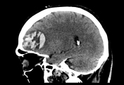
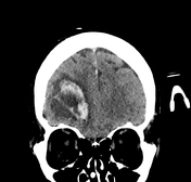
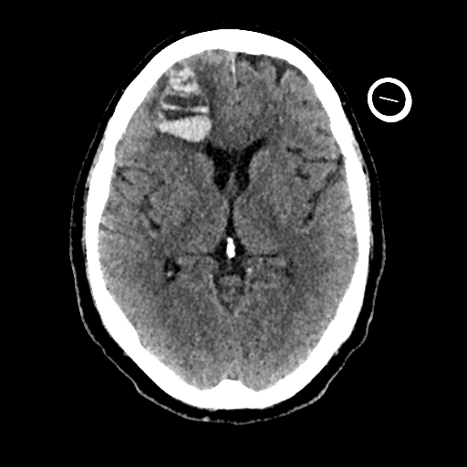
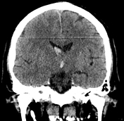
Well-circumscribed intra-axial lesion in the anterior right frontal lobe, which is predominantly markedly hyperdense consistent with acute haemorrhage. It measures 4.6 x 3.8 x 3.6 cm (AP x transverse x craniocaudal). It also contains multiple areas of fluid attenuation (20 HU), and multiple blood/fluid levels are present. Mild perilesional oedema. Mild positive mass effect as shown by effacement of the adjacent sulci, and partial effacement of the frontal horn of the lateral ventricle. The brain parenchyma is otherwise unremarkable. The ventricles are otherwise within normal limits, as are the basal cisterns.
No significant abnormality of the orbits. No significant abnormality of the imaged portions of the paranasal sinuses. Bilateral mastoid effusions without bony destruction, of doubtful clinical significance. No acute bony abnormality or destructive bony lesion.
IMPRESSION: Acute intra-axial haemorrhage in the anterior right frontal lobe associated with mild mass effect. The presence of multiple blood/fluid levels suggests the underlying cause is anticoagulation or coagulopathy. Haemorrhage into a pre-existing cystic tumour could also give this appearance but is considered less likely.

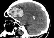
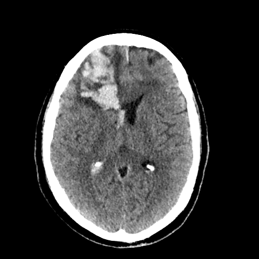
The previously noted intraparenchymal haemorrhage within the right frontal lobe has shown interval increase in size and now measures 3.6 x 5.8 x 4.1 centimetres in maximal transverse, AP, and craniocaudal dimension (previously 3.8 x 4.6 x 3.6 centimetres). There remains surrounding vasogenic oedema with overlying sulcal effacement. There is new effacement of the right lateral ventricle with leftward midline shift of 6 millimetres.
There is now intraventricular extension of the haemorrhage with acute blood in both lateral ventricles (right > left) as well as the third and fourth ventricles. There is no hydrocephalus. The basilar cisterns are maintained. There is trace subarachnoid haemorrhage along the right superior frontal lobe which has developed over the interval. Remainder of the study is unchanged.
IMPRESSION: Interval progression of the known intraparenchymal haemorrhage in the right frontal lobe with new local mass effect, intraventricular extension, as well as new trace subarachnoid blood along the right superior frontal lobe.
Case Discussion
Haemorrhage resulting from anticoagulation can show blood-fluid levels, or haematocrit levels, due to presence of both clotted and unclotted blood. Blood-fluid levels are associated with larger haematoma volume, which is associated with worse clinical outcome. Less commonly, haemorrhagic tumours can display blood-fluid levels.


 Unable to process the form. Check for errors and try again.