
Presentation
Headache, diplopia and vomiting with recent right hemiparesis.
Patient Data
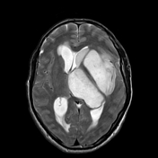


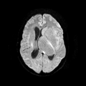
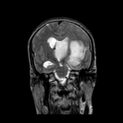
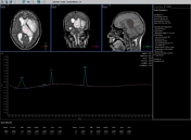

Diffuse involvement of the left insular cortex, medial part of left temporal lobe, left lentiform and caudate nuclei as well as left thalamus with infiltrative process that expands and doesn't distort these structures which display low T1, high T2/FLAIR signal changes. In a previous unavailable post-contrast study there was no enhancement at any of these structures.
The left internal capsule is spared yet there is mass effect upon the lateral ventricles with the ipsilateral ventricle is compressed and contralateral ventricle is dilated with transependymal CSF permeation suggesting subfalcine brain herniation.
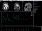
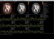
MR spectroscopic data reveals mild Cho elevation, NAA reduction, absence of lactate, elevated Cho/Cr ratio and significant rise of myo-inositol (mI) in intermediate echo in keeping with low grade glioma.
Case Discussion
Biopsy from this lesion revealed WHO grade II fibrillary astrocytoma. Presence of myo-inositol and absence of lactate favour low grade rather than high-grade glioma.


 Unable to process the form. Check for errors and try again.