
Presentation
“Mass” in the left flank for several years. This gets worse with Valsalva maneuver. No history of direct trauma or associated pain.
Patient Data
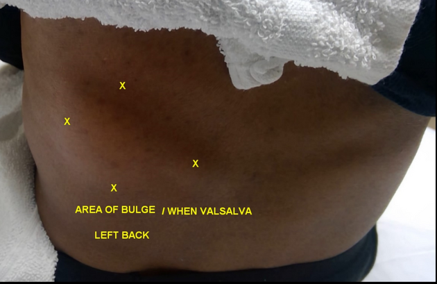
Photo of the patient at the time of ultrasound showing the area of bulge demarcated in the left lower back. The patient is facing away from the camera.


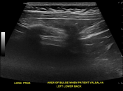
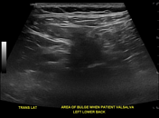
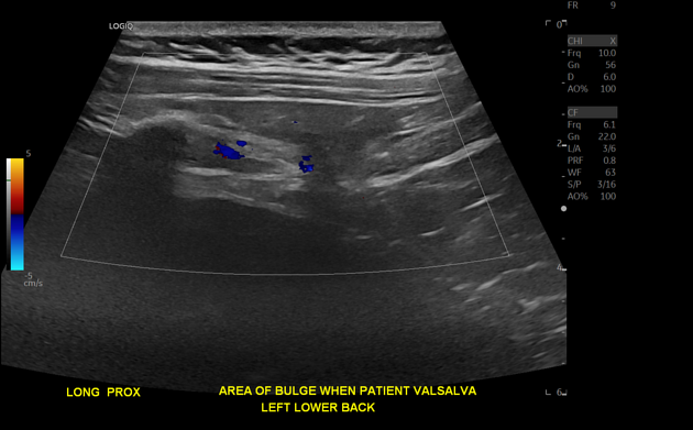
Ultrasound of the left flank shows herniation of fat through a defect in the posterior fascia, which increases with Valsalva maneuver. The neck of the hernia measures approximately 1.1 cm. There is no herniation of bowel loops. The contralateral right side is shown for comparison.
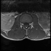
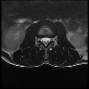

In retrospect, the left superior lumbar hernia was also present on a prior MRI of the lumbar spine, although incompletely imaged at the edge of the field of view on that study. The MRI is from 2 years prior to the ultrasound. The hernia is located posterior to the lower pole of the left kidney. Medial to the hernia is the quadratus lumborum muscle, superior to the hernia is the 12th rib, and superficial to the hernia is the latissimus dorsi muscle.
Case Discussion
This case shows a superior lumbar hernia on ultrasound as well as MRI. If this diagnosis is suspected, Valsalva maneuver should be considered to accentuate it on imaging, as depicted on the cine ultrasound. It is important to exclude bowel herniation on imaging.


 Unable to process the form. Check for errors and try again.