
Presentation
Presented post fall with right wrist tenderness.
Patient Data


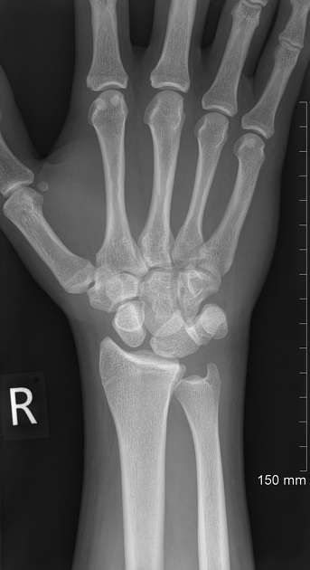
Frontal and oblique radiographs of the right wrist representing typical triangular ( Piece of pie sign (wrist) ) outline of the lunate (please also see annotated images), seen typically in perilunate or lunate dislocation.
Lateral view confirms the diagnosis of lunate dislocation with typical Spilt teacup sign (wrist) .


Annotated frontal and oblique radiographs of the right wrist demonstrating Piece of pie sign (wrist)


Non contrast CT of the right wrist with 3D reformats demonstrates palmar displacement of the lunate bone with disruption of normal radiolunate alignment and evidence of chip fracture of the lunate in its distal aspect.
Additionally 6 mm triangular bony fragment is noted at the dorsal aspect of the wrist suspicious for triquetral bone fracture.


Post K wires fixation images demonstrating anatomical radiolunate alignment.
Case Discussion
This case shows how important it is to look for typical signs of the perilunate or lunate dislocation which is especially difficult on frontal and oblique views. Lateral view should always be obtained if in doubt.
Lunate dislocation should not be confused with perilunate dislocation in which radiolunate articulation is preserved.


 Unable to process the form. Check for errors and try again.