
Presentation
Hemoptysis, chest pain and weight loss.
Patient Data
Age: 60 years
Gender: Female

Download
Info

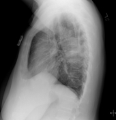
5.9 x 3.6 cm left perihilar mass like opacity.
Mild linear densities in the right lung base suspicious for sub-segmental atelectasis.
No focal consolidation or overt pulmonary edema.
Case Discussion
This represents a case of lung cancer. The patient presented with a 2-month history of chest pain, hemoptysis, and weight loss. A chest x-ray confirmed a left perihilar mass. Biopsy revealed invasive moderately differentiated adenocarcinoma with mucinous features. She underwent systemic chemotherapy following diagnosis confirmation.


 Unable to process the form. Check for errors and try again.