
Presentation
Cough and fever
Patient Data


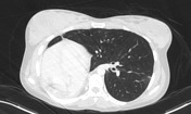
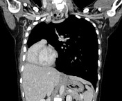
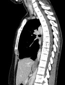
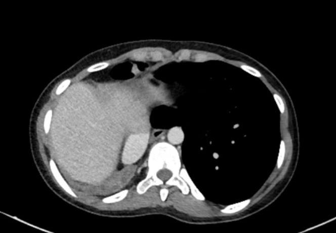
Small collapsed right lung lying posteriorly in the right hemithorax causing cardiomediastinal displacement and rotation. The proximal right main bronchus is seen on the sagittal reformats arising posteriorly and is stenosed and occluded just beyond its origin. Preserved right pulmonary artery and vein. Distal bronchi filled with secretions. Distal focal calcifications. No mass identified.
Compensatory hyperinflation of the left lung, herniated to right side. It shows multiple peribronchial ground-glass opacities and few tree in bud nodules at the anterior segment of left lower lobe suggestive of pulmonary infection.
Left upper lobe pulmonary nodule with mixed densities and foci of calcifications, suggestive of pulmonary hamartoma.
Case Discussion
Features indicate complete chronic collapse of the right lung, an incidental finding in the setting of pulmonary infection. It is one of the causes of opaque hemithorax and volume loss. Asymptomatic cases need no treatment.
The appearance mimics pulmonary hypoplasia, however the right pulmonary artery is similar in size to the left pulmonary artery indicating normal right lung development. This collapse is the result of a chronic acquired proximal bronchostenosis. There is no local calcification, however multiple distal calcific foci in the collapsed lung suggest prior TB.


 Unable to process the form. Check for errors and try again.