
Presentation
2 months postpartum presented with polyuria, dehydration, and seizures. Her laboratory work-up revealed hypokalaemia. Her hormonal profile revealed reduced ADH, FSH & LH levels. Normal ACTH and TSH levels.
Patient Data
Age: 25 years
Gender: Female
From the case:
Lymphocytic hypophysitis


Download
Info
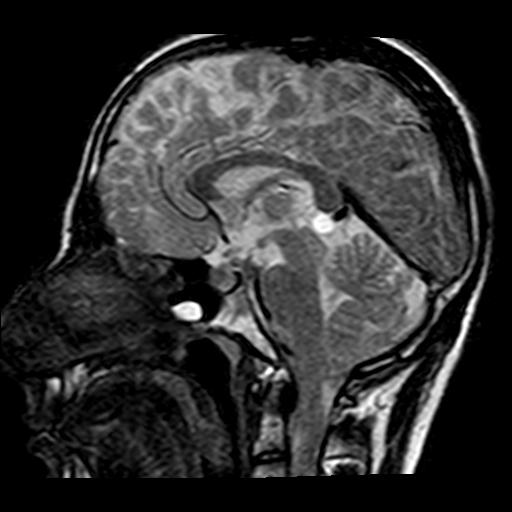
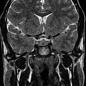
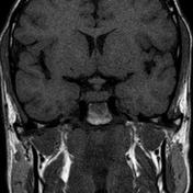
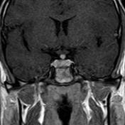
- absent high T1 signal of the posterior pituitary gland
- enlarged avidly enhancing pituitary
- thickened intensely enhancing infundibulum with loss of its inferior tapering.
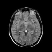
- incidental finding - pineal cyst
Case Discussion
Diagnosis, in this case, is presumptive depending on the clinical, laboratory, and radiological features of the disease. The patient reported a dramatic clinical improvement after corticosteroid treatment.
Radiological features which could differentiate lymphocytic hypophysitis from pituitary adenoma:
- absent posterior pituitary high signal on non-contrast T1 WI
- thickened enhancing pituitary stalk
- bulky pituitary gland showing homogenous enhancement with no focal lesions
- dural tail sign
- parasellar dark signal encasing the pituitary
Differential diagnosis
- pituitary adenoma
- Secondary types of hypophysitis such as granulomatous hypophysitis caused by sarcoidosis, tuberculosis, Wegner, Langerhans cell histocytosis, IgG4 related granulomatosis
- lymphoma
- pituitary metastasis


 Unable to process the form. Check for errors and try again.