
Presentation
Multiple swelling in the foot with discharge.
Patient Data
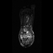

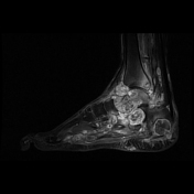
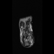
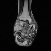
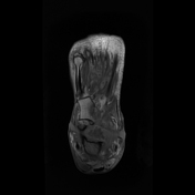
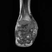
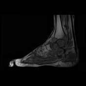
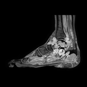

Multiple osteolytic areas are seen in lower end of tibia, talus, calcaneum, tarsal, cuboid, navicular and lateral cuneiform. Osteolytic lesions show multiple discrete and conglomerate rounded lesions showing dot like internal hypointense foci with hyperintense peripheral rim on T1 weighted images. There is extraosseous extension of these lesions in adjacent soft tissue plane.
Multiple discrete and conglomerate small rounded hyperintense lesions with central hypointense dot on T2 weighted images are seen in soft tissue of foot and ankle infiltrating the adjacent muscles. Diffuse oedema and inflammation with multiple pockets of fluid collection, associated discharging sinus tract and skin ulceration is seen. Discharging sinus tracts are more on medial aspect of hindfoot and ankle.
Case Discussion
The characteristic MRI feature is "dot in a circle sign" which appears hypointense dot on both T1 and T2 sequences. Histopathological co-relation and imaging follow-up are not available for this case.


 Unable to process the form. Check for errors and try again.