


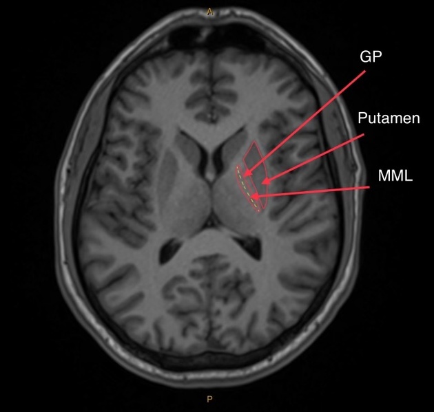
A T1 axial section showing the segments of the basal ganglia, notably the putamen and globus pallidus, with their respective division lines highlighted in yellow.
GP = globus pallidus
MML = medial medullary lamina
Case Discussion
The medial medullary lamina is a bundle of white matter fibres that run between the globus pallidus. Specifically, these fibres delineate the division line between the internal and external segments of the globus pallidus.
Similarly, the lateral medullary lamina divides the external segment of the globus pallidus from the putamen. The internal segment of the globus pallidus can also be subdivided into medial and lateral divisions by a similar structure known as the accessory medullary lamina.
Dysfunction in the globus pallidus, including the medial medullary lamina, may be associated with movement disorders.


 Unable to process the form. Check for errors and try again.