
Presentation
Referred for Tc-per technetate thyroid scintigraphy due to complaints of tachycardia, dyspepsia and chest discomfort. On inspection of neck, there was no asymmetry or nodule. Upon palpation, mild diffuse goitre was palpable. Pulse and respiratory rate were with in normal limits.
Patient Data
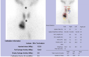

Planar imaging revealed diffuse and homogenous tracer uptake in normal located thyroid gland. An area of increased tracer accumulation was noted in right upper mediastinal region. Thyroid uptake was with in normal range.
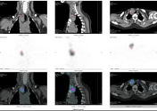
To correctly and surely localise the lesion, SPECT-CT was acquired as well. SPECT-CT revealed soft tissue lesion in superior mediastinum with increased tracer uptake and calcifications.
Case Discussion
This middle-aged female presented to the nuclear medicine department. She was referred for technetium pertechnetate thyroid scintigraphy for suspicion of ectopic thyroid tissue owing to complaints of tachycardia, dyspepsia, and chest discomfort.
An ultrasound of the neck was done at an outside facility, and it showed a nodular swelling originating, most likely, from the thyroid on the right side of the neck.
Pertechnetate thyroid scintigraphy was done 20 minutes after IV administration of 122 MBq of Tc99m pertechnetate. Planar imaging revealed diffuse and homogenous tracer uptake in a normally located thyroid gland. However, increased tracer accumulation was noted in the right upper mediastinal region. Thyroid uptake was within normal range.
SPECT-CT was acquired as well to correctly and accurately localise the lesion. SPECT-CT revealed a soft tissue lesion in the superior mediastinum with increased tracer uptake and calcifications. The patient was advised to undertake thyroid function tests (TFTs) and histopathological correlation.


 Unable to process the form. Check for errors and try again.