
Presentation
Knee pain
Patient Data


There is a triangular lesion within the posterior horn of the medial meniscus that follows marrow signal and has a low signal rim on all sequences. Adjacent to this, no intact root fibres of the posterior horn of the medial meniscus are identified.
The body of the lateral meniscus is mildly enlarged indicating a discoid morphology.
There is moderate delamination of the articular cartilage of the weight-bearing surface of the lateral femoral condyle.


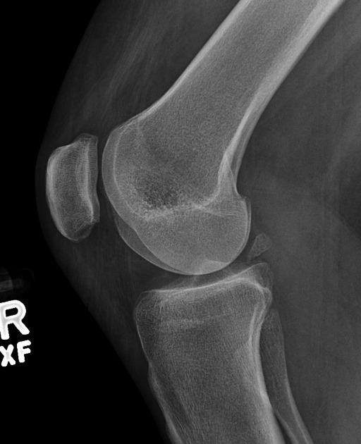
There is a triangular ossific density in the posterior aspect of the medial compartment of the tibiofemoral joint.
Case Discussion
Typical example of a meniscal ossicle, which is a relatively rare ossicle that should not be confused with a fabella, which would be lateral and extra-articular. Meniscal ossicles are often seen in association with, and likely a consequence of meniscal tear, most frequently a root tear.


 Unable to process the form. Check for errors and try again.