
Presentation
Chronic upper abdominal pain.
Patient Data
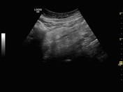


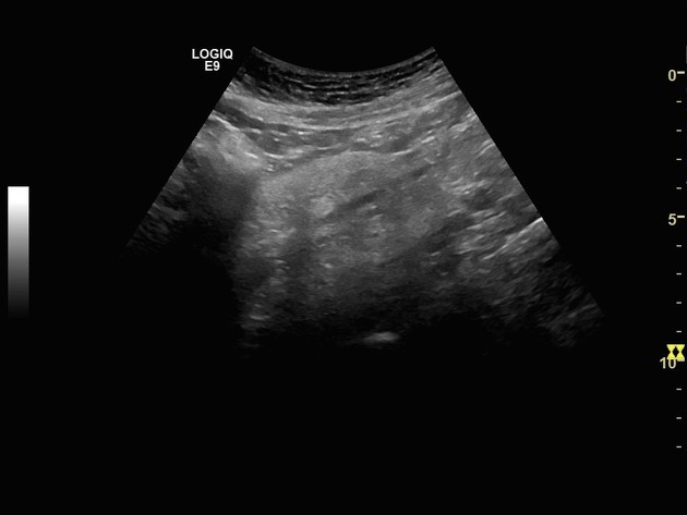
There is hyper-echoic mass-like lesion surrounding the mesenteric vessels in the mid-line of the abdominal cavity.
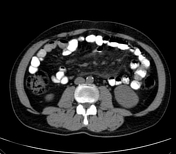
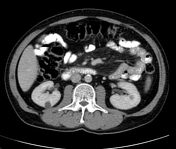
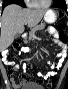
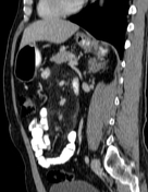

There is a well-defined mesenteric mass-like lesion surrounded by fatty stranding in the root of the superior mesenteric vessels associated with sub-centimetric short-axis mesenteric lymph nodes along with the separation of bowel loops.
Opacification of mesenteric vessels and no abnormal bowel enhancement.
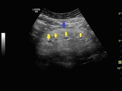
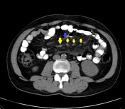
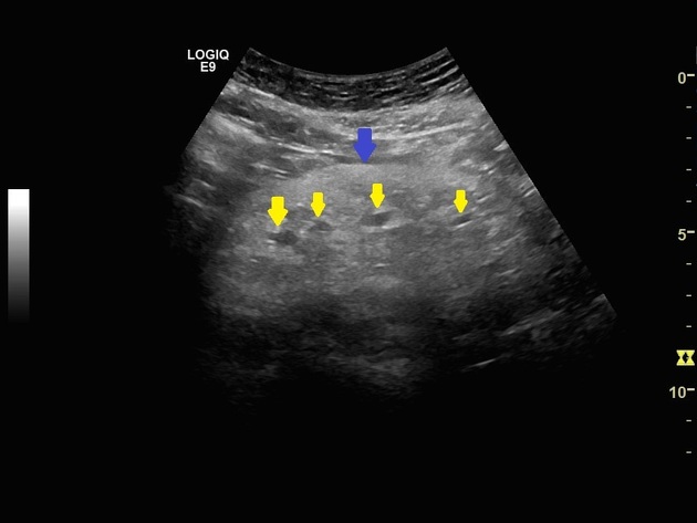
The blue arrow = fatty mesenteric mass-like lesion.
The yellow arrow = intervening mesenteric vessels.
Both are well illustrated in abdominal ultrasound and abdominal enhanced CT scan.
Case Discussion
Mesenteric panniculitis most commonly appears as a fatty mass lesion in the small bowel mesentery with surrounding fatty stranding and mesenteric lymph nodes, it is also non-specific which may be related to malignancy, systemic condition or recent surgery.
It also can be suspected by abdominal ultrasound.


 Unable to process the form. Check for errors and try again.