
Presentation
New-onset left hemiplegia.
Patient Data
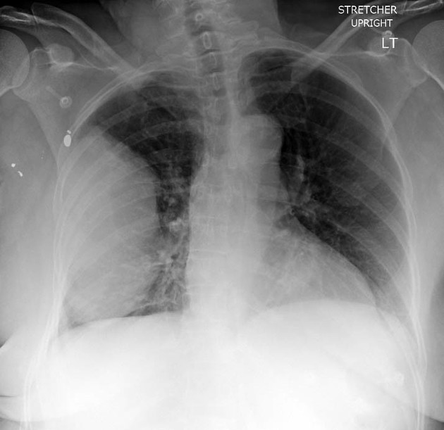
There is a 13-centimetre right lung mass, left lung clear, no pleural effusion. Normal cardiomediastinal silhouette. Status post remote right chest gunshot wound.


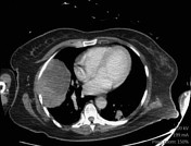
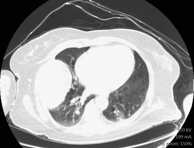
CT chest confirms right lung mass, likely a primary lung cancer given the mild centrilobular emphysema. Mild thickening of the right serratus anterior muscle and enlarged right axillary lymph node are suspicious for chest wall invasion and lymph node spread. Multiple bilateral lung nodules likely represent metastases. No pleural/pericardial effusion, central airways are patent.
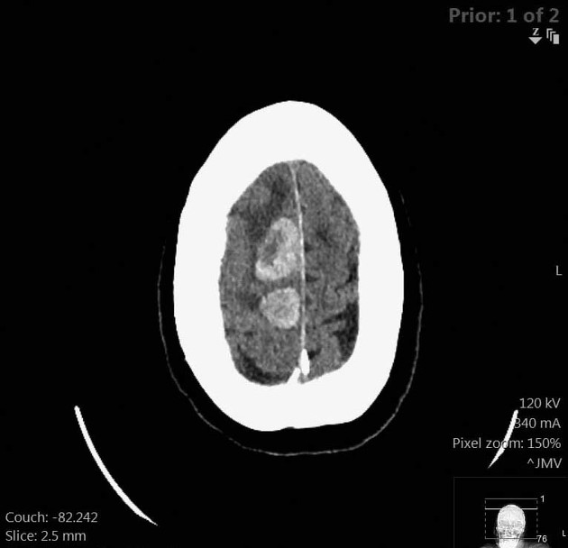
There are multiple intra-axial enhancing/ring-enhancing masses with surrounding vasogenic oedema.
Case Discussion
This patient presented with left hemiplegia, almost certainly due to metastases from previously undiagnosed lung cancer involving the right motor strip. The brain is one of four commonly involved sites for lung cancer metastases. The other three sites are bone, the adrenal glands, and the liver.


 Unable to process the form. Check for errors and try again.