
Presentation
60 pack year smoker, presents with cough and 10kg loss of weight
Patient Data
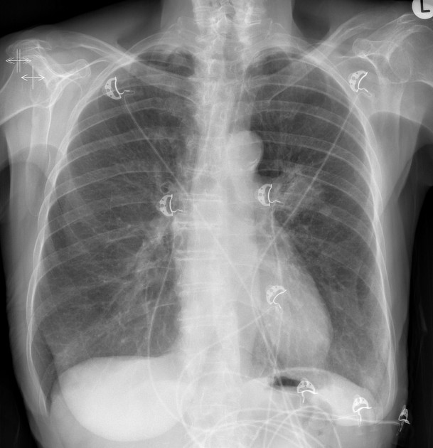
Irregular mass in the left mid-zone appears continuous with lobulated soft tissue density at the left hilum. No surgical clips. Both breast shadows are present.
In a smoker, this is highly suspicious for a primary lung cancer.
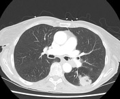

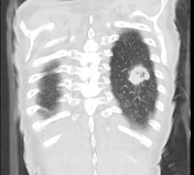
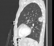
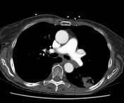
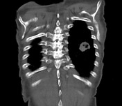
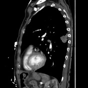
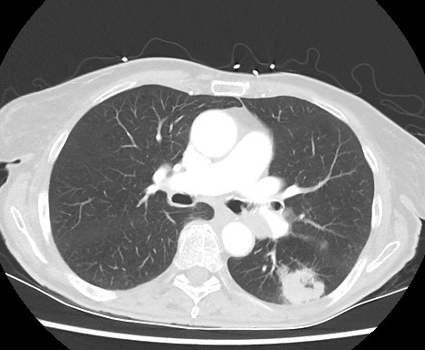
3 cm irregular left lower lobe superior segment mass with tethering of the oblique fissure. 0.8 cm left lower lobe satellite nodule anterior to the larger mass. Left hilar lymphadenopathy. No mediastinal or supraclavicular lymphadenopathy.
The right lung is clear. No pleural effusion.
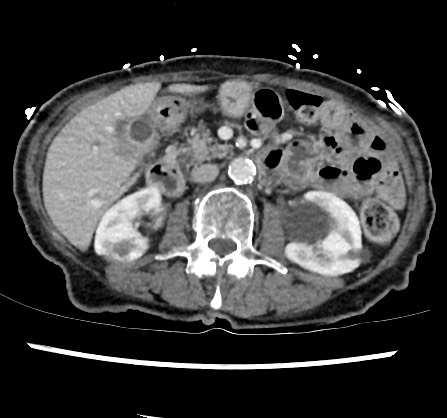
Normal adrenals. Left renal cysts.
Opinion: Left lower lobe primary lung malignancy - T3N1
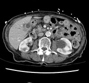
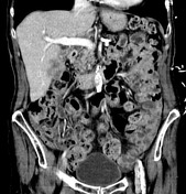
Ill-defined multifocal hypodense lesions in segments 4b and 5 of the liver, adjacent to the gallbladder suspicious for metastases. Small volume pericholecystic fluid and hyperenhancing gallbladder wall.
Normal adrenal glands. No lymphadenopathy. No free fluid. Bilateral renal cysts.
Radiological staging - T3 N1 M1b (Stage IV)
Case Discussion
Liver lesion biopsy: Poorly differentiated lung adenocarcinoma. Epidermal growth factor receptor (eGFR) wild type. ROS1/ALK Immunohistochemical negative. Immunohistochemical PD-L1 0%.
Radiological Staging: T3 N1 M1b (Stage IV).
Conclusion: Stage IV metastatic lung cancer.


 Unable to process the form. Check for errors and try again.