
Presentation
Headache
Patient Data
Age: 75 years
Gender: Male
From the case:
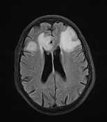
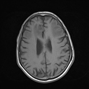
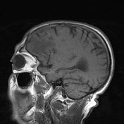
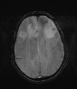
Metastatic lung cancer


Download
Info
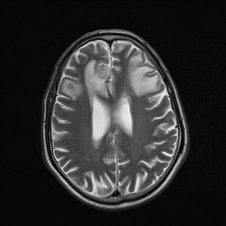
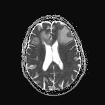
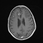
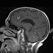
Bilateral frontal, right posterior parietal and right cerebellar ring-enhancing lesions with mild perifocal vasogenic edema and restricted diffusion.
The patient underwent metastatic work-up, starting with CT chest with contrast as a common source of brain metastases.
From the case:
Metastatic lung cancer

Download
Info
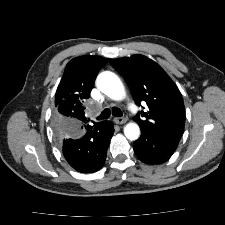
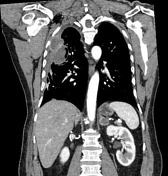
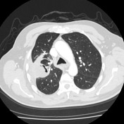
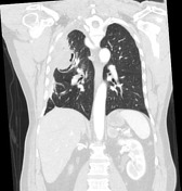
Right upper lobe mass lesion involving the pleura forming a pleural mass lesion with hypodense core and marginal enhancement. It is reaching the mediastinum with ipsilateral hilar adenopathy.
Bilateral adrenal lesions with marginal enhancement, suggestive of bilateral adrenal metastases.
Aberrant right subclavian artery.
Case Discussion
Features are consistent with metastatic lung cancer. It can prersent with brain metastasis.


 Unable to process the form. Check for errors and try again.