
Presentation
Right sided weakness.
Patient Data
Age: 4 years
Gender: Female
From the case:
Moyamoya disease


Download
Info
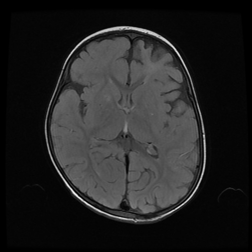
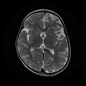
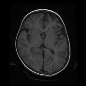
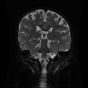
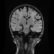
Multiple gliotic areas involving bilateral frontoparietal lobes. Small gliotic areas in bilateral centrum semiovale. Small chronic infarcts in right basal ganglia.
Subtle linear hyperintensities along the sulci bilaterally.
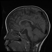
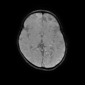
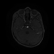
Multiple tortuous flow voids in bilateral basal cisterns, suprasellar cistern and along convexity sulci representing collateral vessels.
Mild asymmetry of lateral ventricles is seen with mild prominence of right
lateral ventricle.
Gross narrowing of supraclinoid segments of both internal carotid arteries with multiple prominent lenticulostriate, thalamostriate, leptomeningeal and dural collaterals.
Case Discussion
Findings are characteristic of moyamoya disease


 Unable to process the form. Check for errors and try again.