
Presentation
5 week history of headache. Now confusion and left foot drop/leg weakness. SOL or CVA?
Patient Data
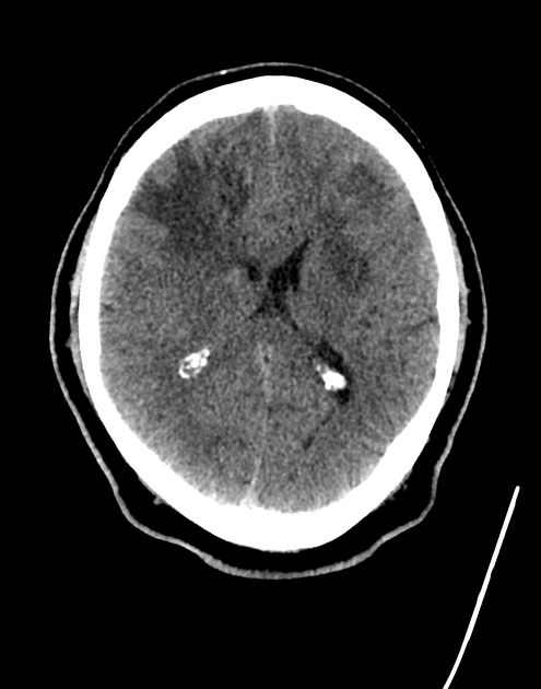
Right frontal lobe mass infiltrating the corpus callosum and crossing the midline.
At least two smaller similar left frontal lesions.
Significant perilesional white matter oedema with respect to both lesions and in the right parietal lobe near the vertex.
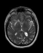

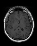
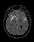
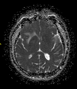
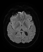
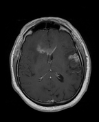
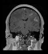

4.9cm right frontal irregular ring enhancing lesion infiltrating the corpus callosum.
1.2cm right parietal and multiple morphologically similar left frontal lesions, the largest 5.6cm.
Significant perilesional white matter oedema.
Case Discussion
This a case of multicentric glioblastoma - there is no communication or altered MR signal between the largest butterfly component and the other lesions in the cerebral hemispheres.
It is therefore the less common multicentric rather than multifocal subtype.


 Unable to process the form. Check for errors and try again.