
Presentation
Stage IVA (pT4pN0) sarcomatoid squamous cell cercinoma. Status post-segmental mandibulectomy, partial maxillectomy, free fibular flap. New bleeding mass on left upper gum/buccal space.
Patient Data


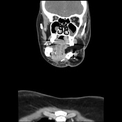
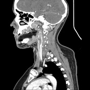
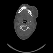
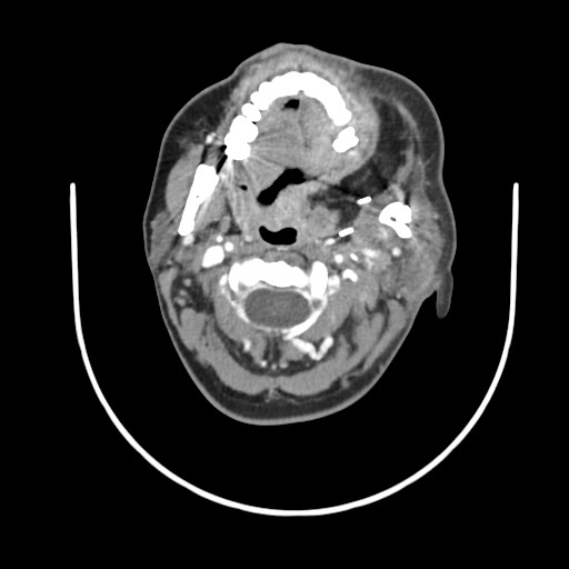
Multifocal new lesions in the oral cavity and oropharynx, including:
13 x 14 mm nodular soft tissue mass at the left gum/buccal space
19 x 16 mm soft tissue mass at the left sublingual/submental region
25 x 13 mm soft tissue mass at the left soft palate
21 x 13 x 27 mm soft tissue nodule at the deep margin of the flap extending superiorly into the medial pterygoid muscle
15 x 13 mm ill-defined soft tissue nodule within the left parapharyngeal space
16 x 16 mm intramuscular soft tissue module in the left infratemporal fossa
11 x 12 mm soft tissue nodule at the left paravertebral muscle at the level of C2
Multiple new punctate enhancing nodules within the left sternocleidomastroid muscle
3 mm nodule anterior to the thyroid isthmus
New nodal changes include:
two left level 2 node with enhancing rim and mild central low hypointensity
Prominent left cervical and supraclavicular nodes are mildly larger compared to previous scans
Case Discussion
This patient initially underwent surgical management with wide-local excision, mandibulectomy, maxillectomy, neck dissection, and free fibular flap, for a histologically proven sarcomatoid poorly differentiated squamous cell carcinoma (SCC) of the retromolar trigon (RMT). The initial post-operative histology demonstrated no nodal metastasis and only local disease.
The patient was referred for definitive post-operative radiotherapy, and the planning CT for the treatment demonstrated no new lesions.
A follow-up scan was organized (above scan) after the patient reported a bleeding mass in the oral mucosa, near the left upper gum/buccal space.
A biopsy of the lesion demonstrated again sarcomatoid SCC, confirming the diagnosis of locoregional recurrence. The patient was referred for systemic therapy.
This case demonstrates the sometimes rapid progression (in this case two weeks) in advance stage, poorly differentiated, oral cavity SCCs.


 Unable to process the form. Check for errors and try again.