
Presentation
New onset tingling in both hands and slurring of speech.
Patient Data


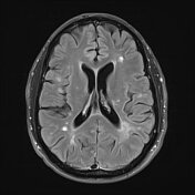
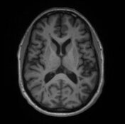
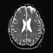
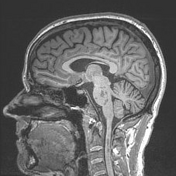
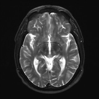
Numerous T2 involving the periventricular, juxtacortical, thalamic, cerebellar, brainstem, spinal cord white matter hyperintense lesions. Several of these lesions are hypointense on T1-weighted imaging.
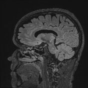
Multiple lesions are orientated perpendicular to the lateral ventricles, best seen on axial T2-FLAIR imaging (Dawson fingers).
Multiple T2 hyperintense lesions have central linear hypodensities, which may represent central vein sign (although no SWI sequences are available to confirm this).
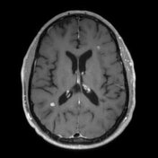
There are numerous enhancing lesions in the supratentorial white matter and at the right pontomesencephalic junction, compatible with active demyelination. A few of these lesions demonstrate incomplete ring enhancement (open ring sign).
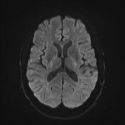
Abnormal enhancement is not seen in the cerebellum or visualised cervical cord. No abnormal diffusion restriction. Brain volume is stable, with overall mild diffuse atrophy.
Case Discussion
This patient's imaging demonstrates classic characteristics of multiple sclerosis (diagnosed formally seven years ago) including perpendicular periventricular lesions (Dawson fingers) and lesions that have incomplete circumferential enhancement (open ring sign). Central vein sign is best seen on T2* / SWI, demonstrated as hyperintense lesions with a thin linear hypointensity that runs completely through the lesion.
This case was submitted with supervision and input from:
Ryan Rigsby, M.D. Neuroradiology Fellow
Mayo Clinic, Jacksonville
Department of Diagnostic and Interventional Radiology


 Unable to process the form. Check for errors and try again.