
Presentation
Right side epiphora and focal soft tissue bulge within medial canthus.
Patient Data


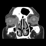
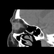
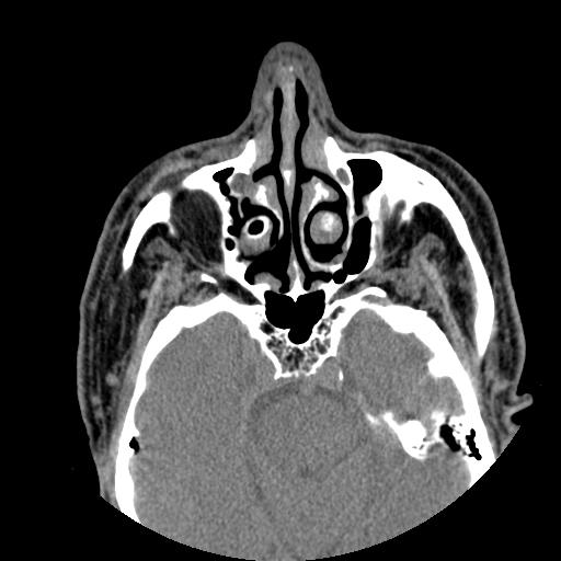
There is a largely cystic right side lacrimal sac and distended right side nasolacrimal duct with the impression of the cystic lacrimal sac on related medial rectus muscle tendon and a uveo-scleral layer of the right eye globe. Note the focal mucosal thickening and mucosal contact within the anterior inferior nasal cavity interposed the adjacent concha and medial wall of the distal nasolacrimal duct.
Case Discussion
The case is a 60-year-old male with a history of right side epiphora and medial canthus soft tissue bulge. The patient has not any history of previous trauma. The orbital and paranasal sinuses non-contrast MDCT requested and obstruction of nasolacrimal drainage apparatus detected by depicting the remarkably enlarged cystic right side lacrimal sac and nasolacrimal canal widening. Dacryocystography in early cases and MDCT in advanced cases are diagnostic.
Dacryostenosis or nasolacrimal duct obstruction (NLDO) is the most common problem of the lacrimal system and can be congenital or acquired. Most of the congenital cases in infants resolve spontaneously by the first year of life. The acquired cases most commonly occur in middle age or elderly females and can lead to acute or chronic dacryocystitis. The pathophysiology of the disorder is not completely known.
The most common symptom is epiphora which can be complicated with infection. The treatment in infants is by massaging or probing the duct and in adults is usually by implanting the stent within the duct or dacryocystorhinostomy.


 Unable to process the form. Check for errors and try again.