
Presentation
Right ear pain and sore throat.
Patient Data
Age: 73
Gender: Female
From the case:
Nasopharyngeal carcinoma

Download
Info

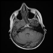
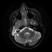
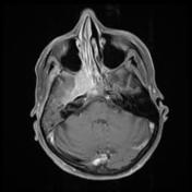
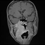
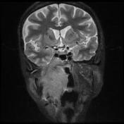
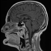
- Right sided nasopharyngeal mass (52x 41x25 mm) involving the right lateral aspect of the oropharynx with subtle mucosal thickening in the posterior wall of the right piriform sinus.
- Extension into the infratemporal masticator space, invading the pterygoid muscles.
- The right internal carotid artery is partially encased.
- Direct invasion into the skull base; greater wing of sphenoid and basi-sphenoid, extending through foramen ovale into the middle cranial fossa.
- Lobulated enhancement is seen along the floor of the middle cranial fossa and the right cavernous sinus is expanded with tumour.
- Right mastoid effusion
- 10mm right jugulodigastric node.
Case Discussion
Case submitted by Dr Smita Deb and A/Prof Pramit Phal.
Large right sided NPC with skull base invasion accounts for the patient's symptoms.
The sites of the nasopharynx include: vault, lateral and posterior walls, superior surface of soft palate.
Staging:
T4- extensive tumour invasion in this case into the middle cranial fossa, infratemporal fossa and masticator space. T4 staging also applies if tumour involves cranial nerves, the brain, hypopharynx or orbit.
N1- unilateral metastasis in lymph node above the supraclavicular fossa <6cm.
The mass was biopsied and histology revealed a well differentiated keratinising type NPC.


 Unable to process the form. Check for errors and try again.