
Presentation
Nasal obstruction for few weeks.
Patient Data
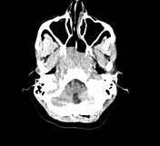

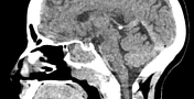
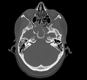

An ill-defined soft tissue density lesion was noted centred in the right nasopharynx, measuring approximately 5.8 x 4.3 x 5.7 cm (CC x AP x trans). There is non-visualisation of the right torus tubarius. A few foci of calcifications are noted within the lesion. No e/o fat attenuation is noted within the lesion. The lesion is extending anteriorly into the posterior portion of bilateral nasal cavities (right > left) via the bilateral posterior choana. The lesion is abutting the bilateral lateral pterygoid muscle laterally. There is e/o mild soft tissue infiltration in the right parapharyngeal space. There is a minimal extension of the lesion into the right pterygopalatine fossa and right sphenopalatine foramen. The lesion is extending into bilateral sphenoid sinuses superiorly and up to the level of the C1 vertebra inferiorly. Posteriorly, there is a loss of fat plane with prevertebral muscles.
There is e/o erosion of the floor of the bilateral sphenoid sinus, the body of the sphenoid, the bilateral greater wing of the sphenoid, petrous segment of the bilateral temporal bone, and clivus.
The bilateral foramen ovale and foramen spinosum are preserved. Mucosal thickening of the bilateral maxillary and ethmoid sinuses is noted.
Ill-defined soft tissue density lesion centred in the right nasopharynx with erosion of the bone of the skull base and extension as described above (D/D nasopharyngeal carcinoma).
Case Discussion
Skull base erosion is seen in stage 3 of nasopharyngeal carcinoma.


 Unable to process the form. Check for errors and try again.