
Presentation
Epistaxis, impaired hearing, and palpable neck lump on the right side.
Patient Data


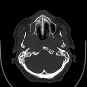
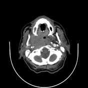
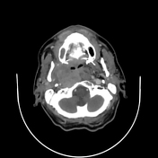
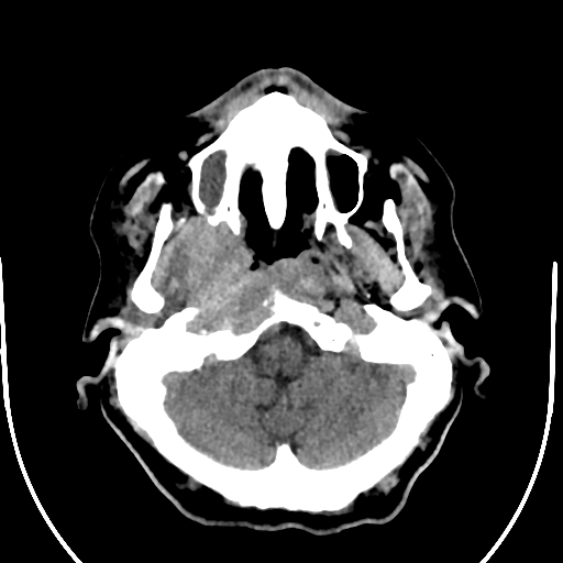
Solid mass measuring 45 x 35 mm with heterogenous enhancement and non-enhancing necrotic areas arises in the right nasopharyngeal mucosa space. The tumour does cross midline to the left nasopharyngeal mucosal space.
The mass infiltrates the retropharyngeal, right sided prevertebral, carotid, parapharyngeal, and masticator space and the corresponding muscles (tensor and levator veli palatini, longus colli and pterygoid).
Torus tubarius, Eustachian tube and the right Rosenmuller fossa are obliterated by the tumour.
Note the bone destruction of the clivus and the right petrous apex.
The mass encases the upper part of the cervical ICA segment as it enters the carotid canal.
Bilateral foramen rotundum, ovale and left foramen lacerum are preserved. Right foramen lacerum are eroded. The tumour encroaches the right hypoglossal canal.
Large right level II lymphadenopathy measuring 40 x 20 mm on the right side and other suspicious rounded shape smaller ones bilaterally above the cricoid cartilage.
Case Discussion
The tumour is highly suspicious for nasopharyngeal carcinoma with skull base erosion.
Staging: According to AJCC 2017, the TNM staging is T3 N2 Mx.
CT is the best modality to evaluate bone destruction as in this case.


 Unable to process the form. Check for errors and try again.