
Presentation
Cough, chronic joint pain.
Patient Data


Multiple pulmonary nodules, some cavitary. Findings are otherwise normal.

CT chest confirms peripheral pulmonary nodules, some cavitary. Findings are otherwise normal.


Lateral views of the cervical spine obtained in the neutral position and extension are normal, but with flexion, the atlanto-dens interval is abnormally wide, indicating that the transverse ligament is torn.

There is a small synovial joint between the back of the dens and the front of the transverse ligament. In rheumatoid arthritis, the synovium may become inflamed, eroding the dens and/or tearing the transverse ligament.
Image credit: Henry Gray (Gray's Anatomy, 20th edition, 1918) 1
Case Discussion
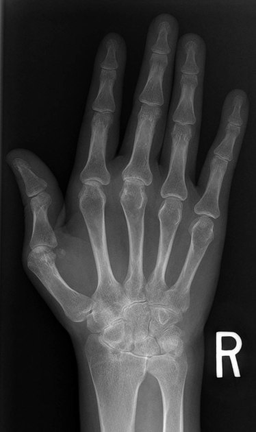
Cavitary lung nodules are most often due to lung metastases (commonly squamous cell primaries) or infections (septic emboli or fungal infections). This patient has characteristic hand (osteopenia and unform MCP/carpal joint space narrowing) and cervical spine (increased atlanto-dens interval with flexion) findings of rheumatoid arthritis, suggesting that the lung nodules are necrobiotic nodules.


 Unable to process the form. Check for errors and try again.