
Presentation
Vaginal delivery. The mother noticed absent right elbow movements.
Patient Data

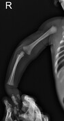
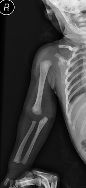
Suspicious abnormal elbow joint alignment in the AP view. The lateral view shows inadequate positioning. No cortical breach.


The symptomatic right side shows a distal humeral metaphyseal fracture with a dorsal angulation at the fracture site. No dislocation.
Cine-loops are aligned to the elbow short-axis on the anterior side.
The asymptomatic side was examined for comparison.
Case Discussion
A mother noticed absent right elbow movement of her week-old baby. The elbow radiographs were negative for a fracture but suspicious for subluxation. The lateral view does not show a lateral position of the distal end of the humerus. Ultrasound was done at the same time and it shows a distal humeral metaphyseal fracture involving the posterior cortex without dislocation.
The case shows a neonatal distal humeral fracture presumed to be a birth injury.


 Unable to process the form. Check for errors and try again.