
Presentation
Preterm neonate with low birth weight and birth asphyxia. Initial routine cranial ultrasound screening at day one showed features of sub-acute meningitis. Now convulsing with reduced oxygen levels.
Patient Data
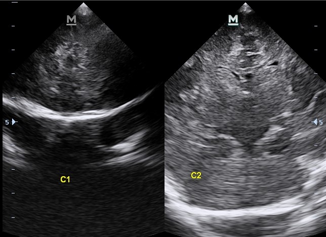
Day 1 findings:
Mild diffuse increased reflectivity and thickening at the leptomeninges and the extra-axial spaces with resultant mildly compressed (slit-like) ventricles are noted. No space-occupying lesions.
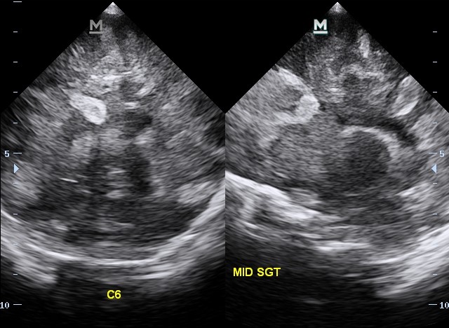
Short interval (worsened) change in three days compared with the initial ultrasound on day one. Widespread cerebral parenchymal oedema with overtly thickened and increased echoicity of the leptomeninges and the extra-axial spaces. A new occurrence of a focal, circumferential echo-dense region at the right periventricular white matter aspect creating a mass effect ipsilaterally and partially extrinsically compressing the right lateral ventricle and mid-line falx cerebri to the left is noted. Spectral Doppler analysis shows absent end diastolic flow within the left posterior communicating artery.
Case Discussion
Features that strongly reflect worsening meningitis and a new occurrence of the unilateral right-sided echo-dense cerebral region which could be cerebritis or attributed to acute haemorrhage owing to its increased reflectivity (grade 1 periventricular leukomalacia) both resulting in increased intracranial pressure. Nevertheless, the neonate improved remarkably with subsequent follow-up ultrasound (not attached) showing unremarkable resolution, on intravenous antibiotic therapy. Lumbar puncture and/or brain computed tomography could not be performed as the neonate was clinically thrombocytopenic and on a ventilator.


 Unable to process the form. Check for errors and try again.