
Presentation
On post-natal clinical examination by a paediatrician, the patient was not irritable but had a right-sided scrotal hard swelling, suspecting a testicular mass. A scrotal ultrasound was requested.
Patient Data

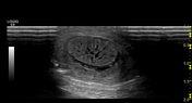
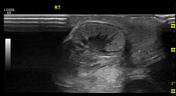
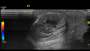

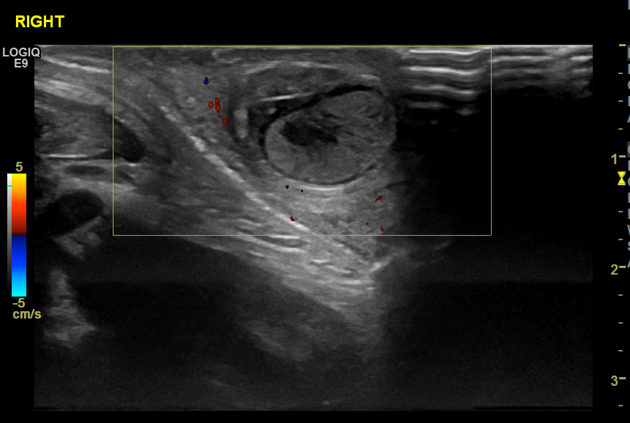
The right testicle appears swollen, has heterogeneous echogenicity, and has hypoechoic linear radiating foci with no detected Doppler colour flow.
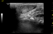
The left testicle shows normal size and echogenicity.
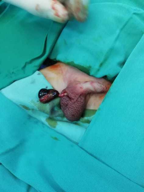
A right testicular excision surgery was done, revealing a mostly black-coloured testicle denoting a non-viable right testicle.
Case Discussion
In suspected testicular torsion cases, the calmness status of the patient on examination (clinical and sonographic) should raise the possibility of irreversible changes that cross over to the more potentially painful acute phase, keeping in mind detorsion probability.
Often, the Doppler colour flow and pulsed wave are difficult to detect in children's normal testicular ultrasound, limiting the diagnosis-dependent features on echo pattern and size change with clinical correlation.


 Unable to process the form. Check for errors and try again.