
Presentation
Seizures following a viral fever.
Patient Data
Age: 20 days
Gender: Female
From the case:
Neonatal viral encephalitis


Show annotations
Download
Info
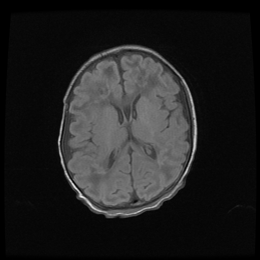
Diffusion restriction is seen involving the corpus callosum and bilateral periventricular/deep white matter. T1 hyperintensities/microhemorryhagic white matter foci are also seen in places which elicit low signal on T2 WI.
Focal microhaemorrhage is seen in the right frontal periventricular white matter.
Case Discussion
The clinical history and pattern of brain involvement are highly suggestive of neonatal viral encephalitis. This can be seen in parechovirus, rotavirus, enterovirus etc. A similar pattern can however also be seen in hypoxic-ischaemic insult. The clinical scenario is the key.


 Unable to process the form. Check for errors and try again.