Neuroblastoma (chest)
Updates to Study Attributes
Large right-sided chest mass with mediastinal shift to the left. ET tube position is appropriate. No bony abnormality. Left lung and right upper lobe (compressed) appear normal.
Updates to Study Attributes
MIBG images. High uptake in the tumour. No metastatic disease. The remainder of uptake is physiological.
Updates to Study Attributes
Reduction in volume of disease following three rounds of chemotherapy.
Image MRI (STIR) ( update )
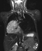
Image MRI (STIR) ( update )

Image MRI (T2) ( update )

Updates to Study Attributes
Huge right-sided tumour within the chest. There is internal calcification.
The tumour extends through neural foramina into the spinal canal although the spinal cord is not compressed.
Small right-sided pleural effusion, sub-pulmonic effusion and trace fluid around the liver.
Image MRI (DWI) ( update )

Updates to Study Attributes
CT confirms internal calcification. No bone destruction (bone windows not uploaded).
Updates to Case Attributes
Appearances here are of neuroblastoma. Neuroblastoma in the chest is unusual, but there are numerous hallmarks of the disease here including a paraspinal location, calcification, neural-foraminal extension and a lobulated appearance. The MIBG update confirms the diagnosis and no metastatic disease.
- +<p>Appearances here are of <a title="Neuroblastoma" href="/articles/neuroblastoma">neuroblastoma</a>. Neuroblastoma in the chest is unusual, but there are numerous hallmarks of the disease here including a paraspinal location, calcification, neural-foraminal extension and a lobulated appearance. The MIBG update confirms the diagnosis and no metastatic disease. </p>
Updates to Freetext Attributes
Pathology
The morphological appearances are those of a neuroblastoma which shows evidence of ganglion cell differentiation (estimated as >5% of the cells) and therefore is classified as a neuroblastoma differentiating. This morphological diagnosis is confirmed on immunohistochemistry with strong positivity with antibodies to CD56 and synaptophysin. Only very occasional cells exhibit positivity with chromogranin. Desmin is negative.