
Presentation
A swelling in the anterolateral aspect of the distal third of the leg. Pain is radiating to the dorsolateral aspect of the foot.
Patient Data
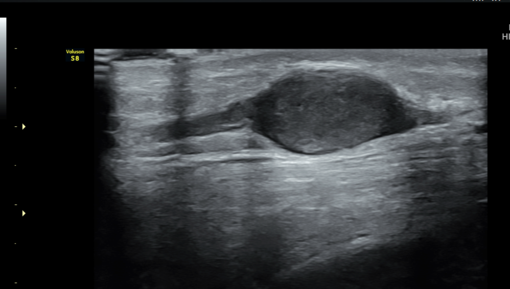
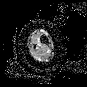
A well-defined fusiform-shaped hypoechoic lesion is noted in the subcutaneous plane of the anterolateral aspect of the distal third of the leg anterior to the peroneus tendon along the course of the superficial peroneal nerve. There is no significant vascularity on the colour Doppler. There is no evidence of invasion into the surrounding structures.
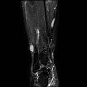

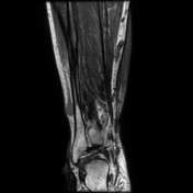
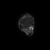
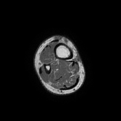
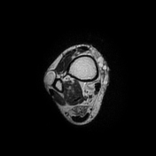
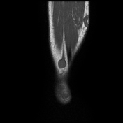
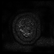

A well-defined oval-shaped T1W isointense, T2W STIR hyperintense lesion with mild restricted diffusion and low ADC is noted along the course of the superficial peroneal nerve in the subcutaneous plane of the anterolateral aspect of the distal third of the leg. The lesion is centred along the nerve with tapered ends showing an 'entry and exit nerve sign'. The nerve can't be separated from the lesion. There is no evidence of cystic changes within the lesion.
Incidentally noted multiple dilated and tortuous superficial varicose veins around the leg.
Case Discussion
The patient presented with a swelling in the anterolateral aspect of the distal third of the leg, with pain radiating to the dorsolateral aspect of the foot. Ultrasound and MRI show features of a benign peripheral nerve sheath tumour along the course of the superficial peroneal nerve.
Neurofibroma and schwannoma are the two most common benign peripheral nerve sheath tumours.
Neurofibroma is predominantly homogeneous and centrally placed along the nerve, whereas schwannoma is eccentrically placed with more heterogeneous cystic degeneration within.


 Unable to process the form. Check for errors and try again.