
Presentation
Memory disorder.
Patient Data


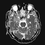


Bitemporomesial signal alterations in T2w / FLAIR and nodular contrast enhancement.
MRI spectroscopy (not shown) was inconclusive.
Case Discussion
The patient went on to have brain biopsy. The biopsy (left mesial temporal lobe) revealed the diagnosis of neurosarcoidosis, it showed predominantly perivascular lymphocellular and plasmacellular infiltrations, reactive astro/microgliosis and diffusely spread noncaseating granulomas consisting of epitheloid cells / histiocytes, immunohistochemistry was typical for sarcoidosis. Tuberculosis was ruled out with PCR.
Cerebrospinal fluid sarcoidosis parameters were elevated (Neopterin 14 (Ref. <1,5), sol. IL2-Receptor 129 IU/ml (Ref. <50), Lysozyme 4,4 mg/l (Ref <1,0), CD4/CD8-Ratio 12 (Ref. <4). ACE was not elevated.
CSF cell count was 40/μl, Total protein was 1991 mg/l. Oligoclonal bands positive. Antineuronal antibodies negative.
Immunosuppressive therapy was initiated (prednisolone, azathioprine), under that therapy the MRI signal changes were slowly regressive but the amnestic syndrome was only partially regressive.


 Unable to process the form. Check for errors and try again.