
Presentation
Patient presents with a recent left frontal mass.
Patient Data


The CT without contrast shows a parenchymal hypodensity in the superior and middle frontal gyrus with subgaleal thickening.
There is also a thickening of the nasal mucosa with bilateral filling of the ethmoidal sinus


The thoraco-abdominopelvic CT shows multiple regular, homogeneous mediastinal, bilateral pulmonary hilar and periportal lymph nodes.
There is no involvement of the pulmonary parenchyma.


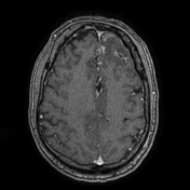


The MRI shows a parenchymal lesion, affecting the cortex and white matter of the superior and middle left frontal gyrus, it is hypointense on T1, hyperintense on T2/FLAIR and slightly hyperintense on DWI without restricted diffusion.
Left frontal parasagittal dural nodular thickening.
There is irregular and nodular lepto-meningeal and subgaleal soft tissue thickening and gadolinium enhancement.
There is nasal and bilateral ethmoidal sinus mucosal thickening.

A nasal biopsy was done, the nasal mocosa is the site of non-caseating granulomas suggesting sarcoidosis.


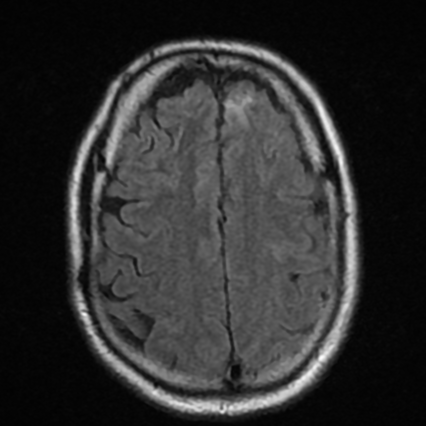
MRI was done six months after corticosteroid therapy, compared with the previous CT, it shows a good response (no nodular lepto-meningeal enhancement, no subgaleal soft tissue lesion, and a regression of the left frontal parenchymal lesion and nasoethmoidal mucosal thickening.
Case Discussion
This case demonstrates stage 1 pulmonary sarcoidosis, and extrapulmonary sarcoidosis (neurosarcoidosis and nasosinosal sarcoidosis), proven by histopathology.


 Unable to process the form. Check for errors and try again.