
Presentation
Incidental finding.
Patient Data
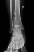
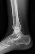

A lytic lesion with sharp lobulated contour and sclerotic margins (narrow transition zone), eccentrically located in the distal tibia, with focal thinning of the adjacent cortex. The lesion measures approximately 32 × 12 × 11 mm.
No pathological fracture, periosteal reaction, associated adjacent soft tissue component is observed.
Case Discussion
The imaging findings are most consistent with an non-ossifying fibroma (NOF). The lesion is greater than 2 cm in size and is therefore classified as a non-ossifying fibroma rather than a fibrous cortical defect.
Non-ossifying fibromas are considered “leave alone” or “do not touch” lesions and are benign, self-limiting. In this case, the lesion was incidentally detected, asymptomatic, and no pathological fracture was observed; therefore, no treatment is required.


 Unable to process the form. Check for errors and try again.