
Presentation
Trauma context
Patient Data
Age: 35 years
Gender: Female


Download
Info

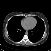
Technique: Volumetric axial images were obtained through the thorax without contrast media injection.
Findings: The lungs and airways are normal. No pleural effusion or thickening. Heart size is normal. No pericardial effusion. The mediastinum structures have normal configuration. Chest wall is unremarkable.
Conclusion: Normal exam.
Case Discussion
This case illustrates a normal chest CT exam, for other examples like this one, please refer to the article on normal chest imaging exams.


 Unable to process the form. Check for errors and try again.