
Presentation
Headache. Pregnant.
Patient Data
Age: 30 years
Gender: Female
From the case:
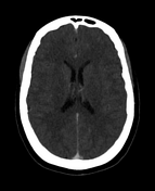
Normal head CT venogram


Download
Info

No hemorrhage, surface collection, mass effect or midline shift. No space-occupying lesion. Grey-white matter differentiation is preserved. The ventricles and basal cisterns are symmetric and normal for age. No filling defects within the dural venous sinuses or cortical veins.
Case Discussion
Case example of a normal head CT venogram (CTV).


 Unable to process the form. Check for errors and try again.