
Presentation
Normal appearance on MRI epilepsy protocol
From the case:
Normal MRI epilepsy protocol


Download
Info
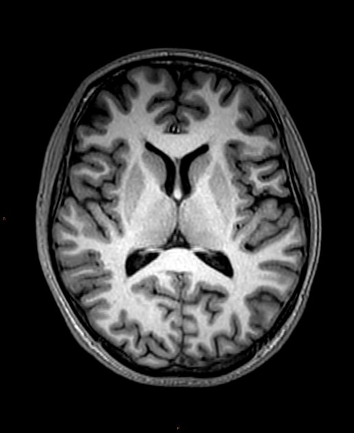
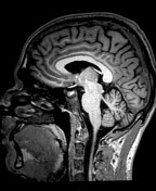
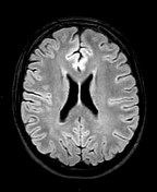
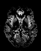
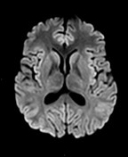
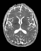
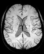
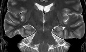
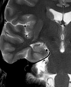
Normal 3.0 T epilepsy protocol:
- volume T1w,
- volume FLAIR (including windowing for cortical abnormalities),
- axial T2w,
- DWI/ADC,
- SWI,
- coronal thin slice T2w (with zoomed bilateral and right temporal lobe views)
From the case:
Normal MRI epilepsy protocol

Download
Info
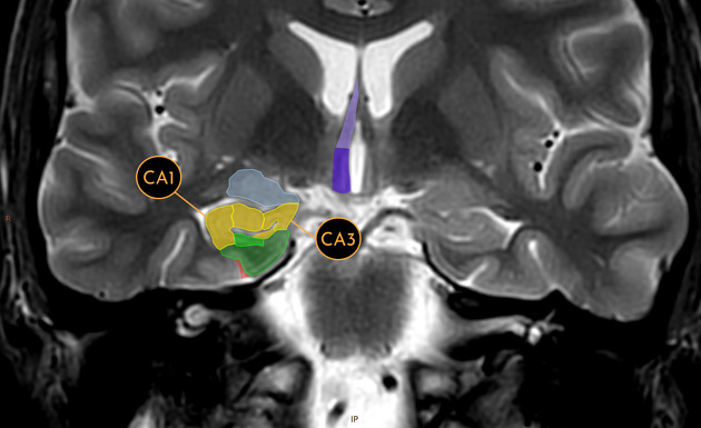
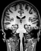
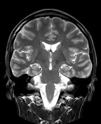
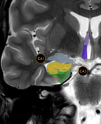
Medial temporal structures (anterior to posterior appearance):
- green - parahippocampal gyrus
- red - collateral sulcus
- blue - amygdala
- yellow - hippocampal head with labeled cornu ammonis 1 and cornus ammonis 3
- purple - mamillary body
- lavender - fornix
- grey - temporal choroid
- orange - hippocampal body / tail
Case Discussion
Annotated images from a normal 3.0 T epilepsy protocol.


 Unable to process the form. Check for errors and try again.