
Presentation
History of clenching and mild pain in the jaw
Patient Data
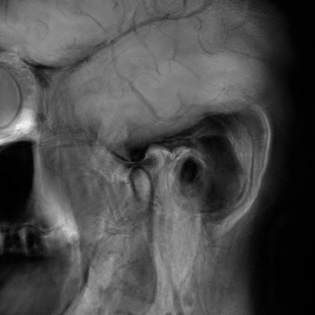
This MRI shows a normal temporomandibular joint with normal function.
Case Discussion
There are different imaging modalities used to evaluate the temporomandibular joint (TMJ) including the US and CT 1.
MRI however, is considered the best imaging modality to make a diagnostic assessment of TMJ abnormalities and status. MRI is the gold standard due to its superior contrast resolution and ability to acquire dynamic imaging for demonstrating the functionality of the joint. MRI can depict the complex anatomy of the TMJ including disc, articular surface, fibrous capsule, ligaments, and muscles of mastication as shown in the example above 2.
Case Courtesy of Dr. Zane Sherif and Mr. Ben Kennedy.


 Unable to process the form. Check for errors and try again.