
Presentation
One year history of a non-productive cough, associated with hoarseness of voice. Ultrasound to rule out goiter and laryngeal structural anomaly.
Patient Data
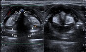


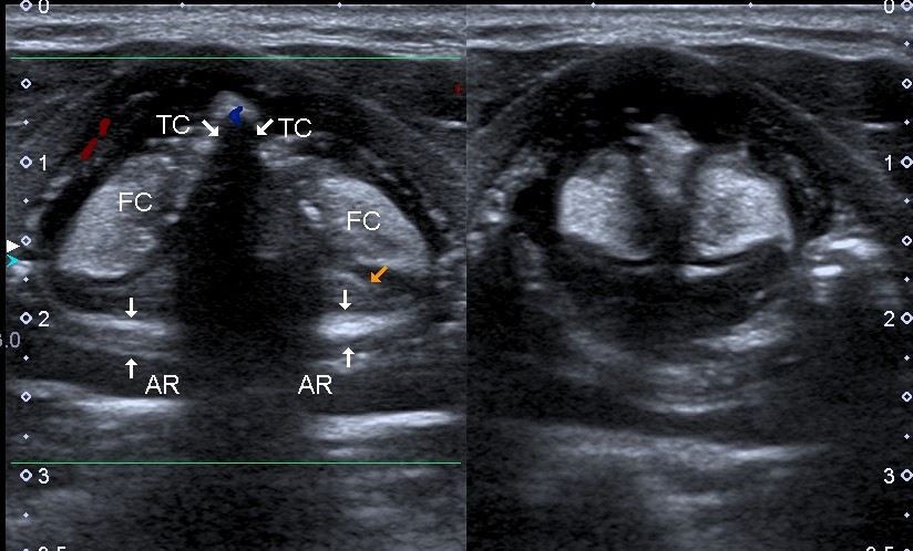
High resolution linear sector array transducer B-grey and color Doppler systematic and dynamic scanning of the larynx at the anterior neck reveals normal laryngeal structures including the true cords (TC), the bilateral false cords (FC), the arytenoid structures (AR), the airway, and the tracheal cartilaginous (C) outline peripherally. No focal space occupying lesions.
Case Discussion
Unremarkable laryngeal structures including the vocal cords and the airway. Notice the lengthening and contractile nature of the cords on abduction and adduction and phonation during real-time ultrasound dynamic assessment. The superior laryngeal artery is visible at the level of the true cords on color flow Doppler mapping.
Transcutaneous laryngeal ultrasonography is readily available, less expensive and utilizes non-ionizing radiation. It is important to assess the mobility of the vocal structures on inspiration and expiration maneouvres during sonographic evaluation. In this illustration, the thyroid gland (images not shown) was normal.


 Unable to process the form. Check for errors and try again.