
Presentation
Bilateral vision loss for 1 month.
Patient Data


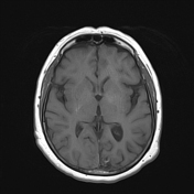
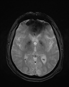
Encephalomalacic changes with surrounding gliosis involving the medial aspect of the left visual cortex. No diffusion restriction.
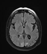
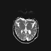
There is a FLAIR high signal involving the medial aspect of the right visual cortex. Faint diffusion restriction is noted in the region. Similar features are also seen in the right side of the splenium of the corpus callosum.
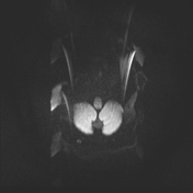
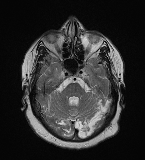
Tiny area of diffusion restriction in the right centrum semi-oval, likely an acute lacunar infarct.
Ex-vacuo prominence of the occipital horn of lateral ventricle -prominent on the left side.
Case Discussion
The features represent subacute and chronic infarcts involving bilateral visual cortexes and the right side of the splenium of the corpus callosum.
Infarctions of different ages can happen due to various factors, such as ongoing vascular disease processes or, most likely, repeated thromboembolic events affecting an area of the brain that has already experienced some degree of ischemic injury.
Several differential diagnoses can cause bilateral occipital or posterior circulation infarcts, including:
atherosclerotic changes of basilar artery
small vessel disease affecting the perforating branches of the posterior cerebral arteries
hypoperfusion (such as in hypotensive states)
cerebral venous sinus thrombosis
migraine with aura (albeit uncommon); some cases of migraine with aura can mimic the appearance of occipital infarcts on imaging studies
No definite cause has been identified in this patient.


 Unable to process the form. Check for errors and try again.