
Presentation
Right ptosis
Patient Data


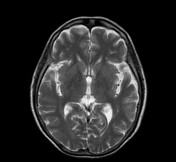
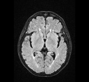
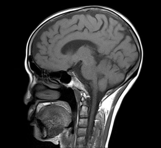
The white matter T2 hyperintensity is patchy in both cerebral hemispheres suggesting chronic small vessel ischaemia.
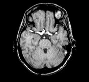
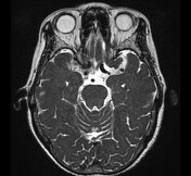
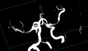
The saccular aneurysm arises from the cavernous part of the right internal carotid artery that compresses into the right oculomotor nerve.
There are no abnormal intensity on the midbrain and no neoplasms ( pituitary macroadenomas, meningiomas, schwannomas) along the course of the right oculomotor nerve.
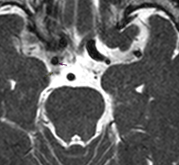
Yellow arrow: right oculomotor nerve
Pink arrow: aneurysm
Red circle: the close relationship between the right oculomotor nerve and the aneurysm.
Case Discussion
MRI shows the right oculomotor contact with an aneurysm of ICA. The imaging findings in a patient with acute ptosis are in keeping with oculomotor nerve palsy due to a carotid aneurysm.


 Unable to process the form. Check for errors and try again.