
Presentation
Dysphagia for work-up. Background weight loss, positive ethanol and smoking history.
Patient Data
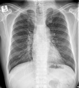
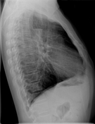

There is a gas-fluid level within the thoracic esophagus with a widened right paratracheal stripe.
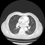
Hyperinflated lung fields are consistent with a history of smoking and COAD.
There are no suspicious nodules or masses.
The left clavicular companion shadow is notably absent.


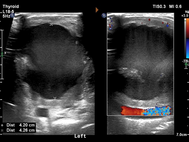
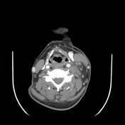
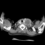
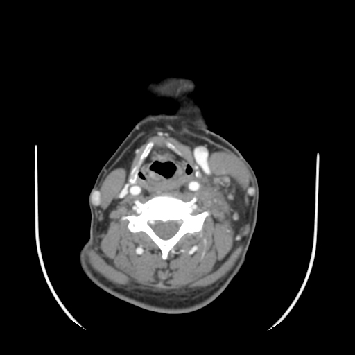
A large, necrotic, left supraclavicular lymph node was identified clinically and targeted for ultrasound-guided FNA. This confirmed metastatic squamous cell carcinoma.
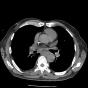

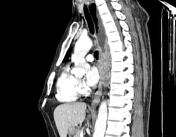
There is a dilated fluid and debris-filled esophagus proximal to an ulcerating esophageal mass at the T8 level (middle third ) measuring at least 32 x 30mm in axial dimensions. The craniocaudal extent is difficult to accurately assess, however, a minimum length of 80 mm is measured.
Features suggestive of esophageal carcinoma of squamous cell origin in view of positive left supraclavicular lymph node FNA.
There is no invasion of the adjacent aorta or left atrium. There is mild right-sided tracheal displacement, however no tracheal invasion. There are no pulmonary or osseous metastases.
Multiple necrotic lymph nodes are demonstrated:
left supraclavicular lymph node - 54 x 48mm
right level IV lymph node- 17 x 12mm.
left para-esophageal lymph node at the T9 level- 15 x 13mm.
celiac axis lymph node in the upper abdomen - 25 x 21mm.
There is a right subclavian central venous line.
Case Discussion
A case of esophageal carcinoma with metastatic and necrotic lymph adenopathy (provisional stage III- T3, N2, cM0).
The left clavicular companion shadow is absent. This may be normal or due to regional pathology.
In this instance, a large, necrotic, left supraclavicular lymph node was clinically present, confirmed on ultrasound. A guided FNA revealed metastatic squamous cell carcinoma.
A chest X-ray, CT neck and chest were requested for review and initial staging.


 Unable to process the form. Check for errors and try again.