
Presentation
Headache
Patient Data
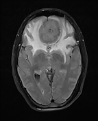

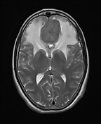
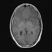
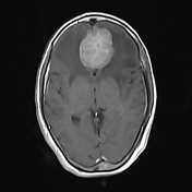
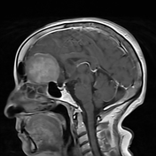

There is evidence of well defined homogenous extra-axial anterior cranial fossa soft tissue mass arising from the olfactory groove and surrounding the falx anteriorly. The lesion exhibiting T1 isointense, T2 hyperintense, vividly contrast enhancing, diffusion restricting. Numerous small flow voids representing vessels are seen around the periphery of the lesion. The mass exerts significant local mass effect with cortical buckling and lateral displacement of the medial aspects of the frontal lobes and posterior displacement of the genu of the corpus callosum. The frontal horns of the lateral ventricles are splayed by the mass and the anterior cerebral arteries are displaced posteriorly and draped over the posterosuperior aspect of the mass.
Moderate surrounding T2/flair signal hyperintensity within frontal lobes and anterior temporal poles bilaterally representing vasogenic oedema. The basal cisterns remain uneffaced. No uncal or transtentorial herniation.
Case Discussion
This case demonstrates typical appearances of an olfactory groove meningioma.


 Unable to process the form. Check for errors and try again.