

Presentation
Visual issues. Progressive. Chiasmal lesion?
Patient Data


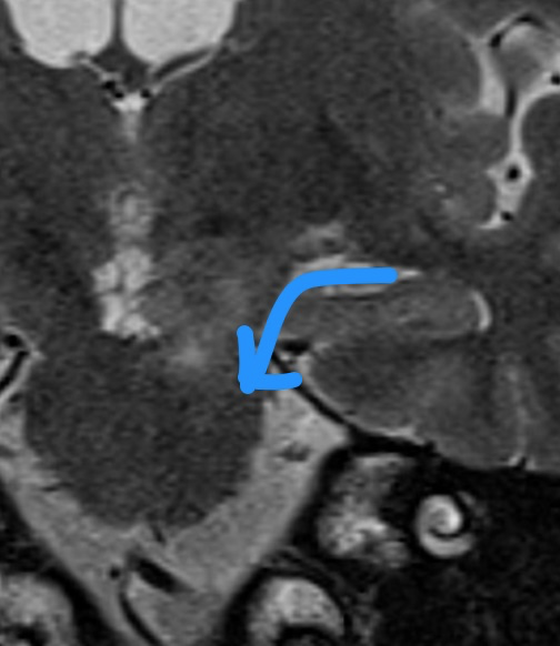
Relatively well defined T1 isointense, T2 hyperintense, avidly and homogeneously enhancing mass in the supra-sellar region, separate from the pituitary gland. The mass is morphologically in keeping with an expansile abnormality in the optic chiasm and the origins of the optic radiations. On the left, it appears to involve the lateral geniculate ganglion.
The optic nerves are normal in appearance. No intra-conal or extra-conal abnormality.
Additional identical enhancing lesion in the left cerebral peduncle and enhancement of the left abducens nerve.
The appearances were thought to be a single lesion (kind of looks like two) linked to the lateral geniculate, which is anatomically confining and hence the odd morphology of the lesion.
Case Discussion
This case resulted in considerable clinical and radiological discussion as to the potential differential diagnoses.
This ranged from demyelinating disease to metastases to an optic glioma.
The patient's visual loss temporarily responded to pulsed steroids.
Later the patient had a surgical biopsy performed at another institution which showed this to represent a B-cell lymphoma.
This is a very good grey-case or 'test the expert' case for educational meeting discussion. This was made all the more difficult as the initial studies were for a para/sellar mass and hence received an MR protocol for pituitary imaging. No DWI-ADC imaging was available which may have helped in the diagnostic process with lymphoma being a highly cellular tumour and hence showing diffusion restriction.
This is a rare site for a primary CNS lymphoma.


 Unable to process the form. Check for errors and try again.