
Presentation
Long-standing Graves disease and performed decompression surgery.
Patient Data
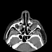

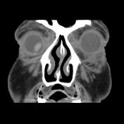
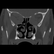
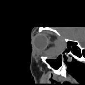

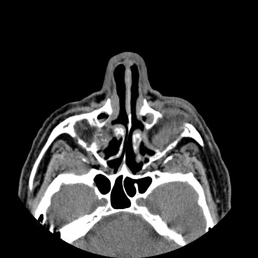
Atrophic lacrimal glands with prolapse, bilateral Proptosis, increased orbital fat, large width extraocular muscles more severe in medial and inferior and superior rectus muscles in both sides and right side lateral rectus muscle, preseptal swelling, left eye no native lens, and inferior-nasal deviation of the right eye is seen.
Large bone defects due to orbitotomy on the orbital medial wall, lateral wall, and floor with related fat and extraocular muscles herniation through the defects are also seen. Foci of thick mucosa in paranasal sinuses more in the right maxillary sinus are noted.
Case Discussion
The case illustrates the typical non-contrast MDCT features of the Graves ophthalmopathy and orbitotomy decompression surgery for the treatment of optic nerve compression due to enlarged extraocular muscles and apical crowding.


 Unable to process the form. Check for errors and try again.