
Presentation
Ongoing left knee nocturnal pain responding to anti-inflammatory drugs
Patient Data
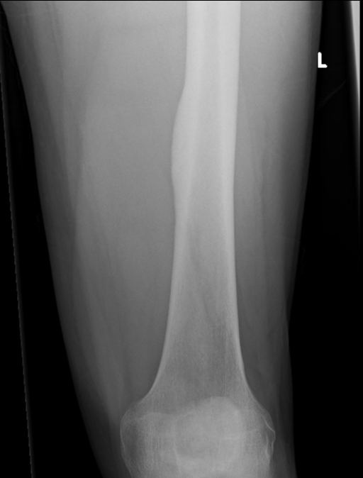
There is thickening of the cortex medially at the level of the mid shaft of the femur.
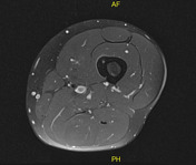

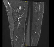
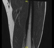

A small nidus of abnormal signal is seen involving the medial cortex of diaphysis of the left femur associated with exuberant cortical thickening. This focus elicits an intermediate signal on T1-weighted images and high signal on fluid-sensitive images. The focus measures 3 mm in its axial diameters and 6 mm in its craniocaudal diameter.
Case Discussion
Findings are consistent with osteoid osteoma.
Osteoid osteoma is a painful and benign bone tumour that is common in young adults. The typical clinical presentation consists of pain that becomes worse at night and is relieved by anti-inflammatory drugs.
Typical locations include intracortical bone and diaphyses of long bones.
When the lesion is located in typical locations (as in our case), both characteristic clinical and radiological features are diagnostic.


 Unable to process the form. Check for errors and try again.