
Presentation
Tender, swollen second toe without trauma.
Patient Data

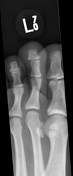
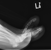
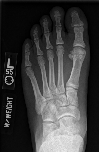
Radiographs of the left second toe demonstrate a well-circumscribed lucent lesion in the distal phalanx.
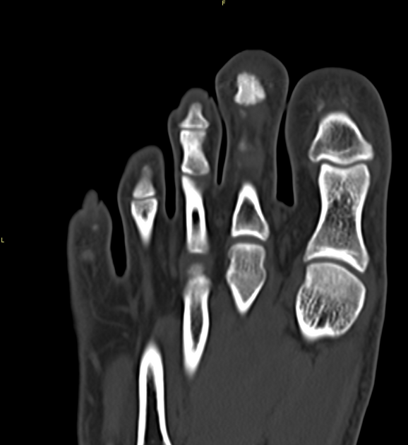
Coronal images from non-contrast CT confirm a lucent lesion with a thin sclerotic margin.
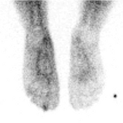


Bone scintigraphy performed using 20 mCi of Tc-99m MDP IV
Blood pool phase obtained 2-10 minutes after injection shows focal increased uptake at the tip of the second toe, left foot.
The delayed phase obtained 3 hours after injection shows focal intense increased uptake at the distal phalanx, second toe, and left foot.


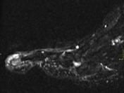
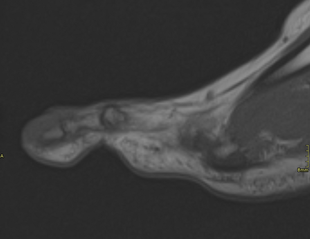
Sagittal T1 and STIR-weighted sequences from MRI show abnormal signal intensity in the distal phalanx and surrounding soft tissues of the second toe.
Hypointense soft tissue on T1.
Hyperintense soft tissue and marrow on STIR.
Post-contrast images were not available due to technical difficulties.
Patient was treated successfully with resection of the phalanx.
Pathologic evaluation showed findings consistent with an osteoid osteoma.
Case Discussion
Osteoid osteomas (OO) are a benign osteoblastic tumour of bone. They are seen most frequently in males during their second and third decades of life. When encountered, OOs most typically involve the diaphysis or metaphysis of long tubular bones such as the femur or tibia and are rarely encountered in the phalanges of the hands and feet.
Radiography: sclerotic cortically based lesion which contains a small lucency that represents the tumour nidus. Less frequently they are medullary based or subperiosteal.
CT: shows similar findings with improved detection of the nidus which may be difficult to see on x-ray.
Bone scan: the classic scintigraphic finding is the “double-density sign”. This is a central focus of intense radiotracer uptake representing the osteoblastic activity of the nidus. Surrounding this is a larger, less-intense intense ring of radiotracer uptake, representing the normal bone reactive changes. This findings is seen mainly in long bones and less frequently in spinal OOs and those found in small bones such as this case.
MRI: while variable, most OOs exhibit hypo-to-isointense signal on T1-weighted images and hyper intense signal on T2-weighted and STIR images. Enhancement may be diffuse or marginal.


 Unable to process the form. Check for errors and try again.