
Presentation
Pelvic pain.
Patient Data
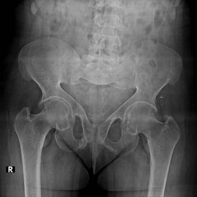
triradiate appearance of the pelvis with bilateral protrusio acetabuli
osteopenic texture of the examined bones with coarse trabeculae
right superior and inferior pubic rami Looser's zones
subchondral bone absorption of sacroiliac joints (more on the right)


MRI exam of the pelvis showed:
triradiate appearance of the pelvis with bilateral protrusio acetabuli
right superior and inferior pubic rami Looser's zones. Also note the marrow oedema of the left femoral neck, likely stress fracture
subchondral bone absorption of sacroiliac joints (more on the right)


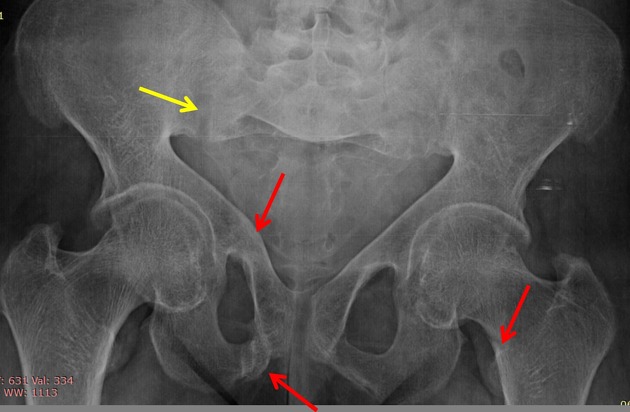
Red arrows: represent stress fractures (looser's zones).
Yellow arrows: represent subchondral bony resorption at the sacroiliac joint.
Case Discussion
The patient had a history of chronic renal disease.
Osteomalacia is bone softening due to insufficient mineralisation of the osteoid secondary to any process that results in vitamin D deficiency or defects in phosphate metabolism.
Radiographic features (musculoskeletal)
There can be variable appearances dependant on the cause :
diffuse demineralisation: osteoporotic-like pattern
coarsened trabeculae
pseudofractures (Looser zones)
-
articular manifestations (uncommon)
rheumatoid arthritis-like changes
osteogenic synovitis
ankylosing spondylitis-like changes


 Unable to process the form. Check for errors and try again.