
Presentation
Diabetic foot.
Patient Data


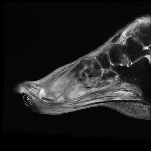
extensive bone marrow signal changes (T1 hypointense, STIR, and PD hyperintense) involving the 2nd and 3rd metatarsal head and neck and proximal phalanx base and shaft compatible with osteomyelitis
medially dislocation of the second toe at the level of the MTP joint
moderate joint effusion at the 3rd metatarsophalangeal joint
sinus tract starts from the level of the 2nd and 3rd metatarsophalangeal joint into the skin in the plantar aspect
amputation of great toe and distal half of first metatarsal
abnormal bone marrow signal (iso on T1 and high on STIR/PD fs) at the first metatarsal mid to proximal diaphysis and medial cuneiform bone due to osteitis
extensive oedema and fluid signal intensity in intrinsic muscles and subcutaneous tissues of the forefoot and midfoot
Case Discussion
Diabetes mellitus can involve the foot through two mechanisms:
The first one is osteomyelitis which mainly affects the pressure points in the forefoot and hindfoot.
The second one is a neuropathic joint which affects the intertarsal joints causing joint destruction, disorganisation, and dislocation.


 Unable to process the form. Check for errors and try again.