
Presentation
Lower abdominal pain. Abdominal ultrasound suggests a haemorrhagic cystic tumour in the left ovary.
Patient Data


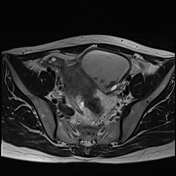
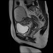
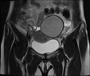
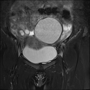
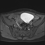
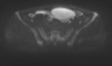
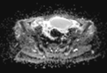
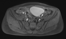
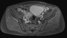
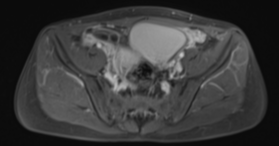
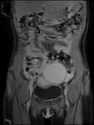
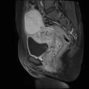
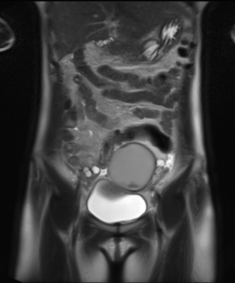
A well-defined, unilocular left ovarian cystic mass (measuring 66 × 47 × 69 mm) demonstrates high signal intensity on T1FS with a shading sign on T2W and contains several nodules with low signal intensity on T2-weighted images ("T2 dark spot sign") with diffusion restriction. No mural nodules, septations, fat, or solid components are observed. No post-contrast enhancement is noted.
Additionally, a uterine fibroid and several functional ovarian cysts bilaterally are also observed.
Case Discussion
The MRI findings are consistent with a left ovarian endometrioma, characterised by the T2 dark spot sign and shading sign. This lesion can also be classified as O-RADS 2.


 Unable to process the form. Check for errors and try again.