
Presentation
Referred for swelling of the head.
Patient Data

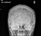
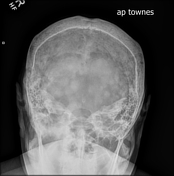
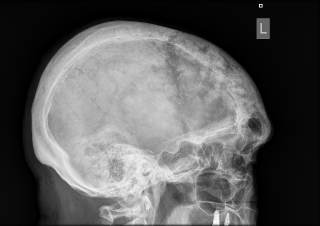
Widening of the inner and outer table most prominently noted over the frontal and parietal region with similar but less severe involvement in the remainder of the cranial bones. Anteriorly there are patchy areas of sclerosis consistent with a "cotton wool" appearance. Additionally, an edge of osteolysis can be seen involving the anterior one third of the skull, consistent with "osteoporosis circumscripta".
Case Discussion
The radiographic findings represent the skull manifestations of Paget disease. There are three primary radiographic stages of Paget disease:
1: Active osteolytic phase: Consists of bony resorption, which has different named appearances based on location. In the skull - osteoporosis circumscripta. While in a long bone - blade of grass or flame like osteolysis.
2: Osteolytic/osteosclerotic: Patchy areas of sclerosis intermixed with the areas of osteolysis.
3: Inactive phase: Thickening of the cranium/cortex with coarse trabeculae. The trabecular pattern creates areas of sclerosis giving the "cotton wool" appearance in the skull.


 Unable to process the form. Check for errors and try again.